A kidney stone can cause pain that begins suddenly and feels impossible to ignore. While kidney stone treatment often starts with controlling pain and helping the stone pass naturally, the right approach depends on the stone’s size, location, symptoms, and whether it is blocking the urinary tract. A urology evaluation can clarify what is happening and help prevent complications.
Kidney stones are hard deposits that form when certain minerals and salts become concentrated in the urine. They may remain in the kidney without causing symptoms, but pain can begin when a stone moves into the ureter, the narrow tube that carries urine from the kidney to the bladder.
Treatment is not the same for every patient. A small stone may pass with fluids, prescribed medication, and time. A larger stone, a stone causing persistent obstruction, or one associated with infection may need a procedure. The goal is to relieve symptoms safely, protect kidney function, clear the stone when necessary, and reduce the chance of another episode.
Many small stones can pass on their own, particularly if they have moved lower in the ureter. A clinician may recommend drinking an appropriate amount of water, taking pain medication as directed, and using medication that relaxes the ureter to make passage easier in selected cases.
Patients may be asked to strain their urine to collect the stone if it passes. Stone analysis can be useful because it identifies what the stone is made of and helps guide prevention. Passing a stone can take days or sometimes several weeks, so follow-up matters. Pain that improves does not always mean the stone has passed completely.
Drinking excessive water during severe pain or nausea is not always helpful and may worsen discomfort. The right fluid intake should be discussed with a clinician, especially for people with heart, kidney, or other chronic medical conditions.
Pain from a moving kidney stone can be intense and may come in waves. Anti-inflammatory medicines are commonly used when medically appropriate because they can reduce pain and swelling around the ureter. Other pain medicines or anti-nausea treatment may also be considered depending on the patient’s health history and symptoms.
Do not rely on over-the-counter pain medicine alone if pain is severe, recurring, or accompanied by other warning signs. A stone can obstruct urine flow, and symptoms that seem manageable at home may still require imaging and medical assessment.
A kidney stone should be assessed urgently when there is fever, chills, vomiting that prevents fluids from staying down, severe or uncontrolled pain, very little urine output, or visible blood in the urine with worsening symptoms. Fever or chills can indicate a urinary infection. When infection occurs behind a blocked ureter, it can become serious quickly and may require emergency drainage and antibiotics.
Pregnant patients, people with one functioning kidney, those with known kidney disease, and people with weakened immune systems should seek timely medical advice when symptoms suggest a stone. The same applies when pain is new and unexplained, since other conditions can cause similar symptoms.
A clinician may use urine testing, blood tests, ultrasound, or CT imaging to confirm a stone, estimate its size, identify blockage, and check for infection. These results help determine whether observation is reasonable or whether treatment should move forward.
When a stone is unlikely to pass, causes ongoing blockage, or leads to repeated pain or infection, a urologist may recommend a procedure. The choice depends on the size and position of the stone, its composition when known, anatomy, and the patient’s overall health.
Shock wave lithotripsy uses focused sound waves from outside the body to break a stone into smaller pieces that can pass in the urine. It may be suitable for certain kidney or upper ureter stones. It is less invasive than surgery, but it is not ideal for every stone type or location, and some patients need more than one session or additional treatment afterward.
During ureteroscopy, a urologist passes a small instrument through the urinary tract to locate the stone. A laser can break the stone into smaller fragments, which may be removed or left to pass. A temporary ureteral stent may be placed afterward to support drainage while swelling settles.
A stent can cause temporary urinary urgency, discomfort, or blood in the urine. Patients should be told when it needs to be removed, because it is not intended to remain in place indefinitely.
For very large, complex, or difficult-to-reach kidney stones, percutaneous nephrolithotomy may be considered. This procedure removes stones through a small incision in the back. It is more invasive than shock wave treatment or ureteroscopy, but it can be the most effective option for larger stone burdens.
Each procedure has benefits and possible risks, including discomfort, bleeding, infection, and the possibility that additional treatment may be needed. A specialist can explain the expected recovery and which option best fits the individual situation.
After a first stone, prevention should be part of the conversation. The most useful plan depends on the type of stone, dietary habits, medications, medical conditions, and laboratory findings. A general recommendation that helps many people is to drink enough fluid throughout the day to keep urine pale yellow, unless a clinician has advised fluid limits.
For many calcium-based stones, normal dietary calcium intake is usually preferred over avoiding calcium completely. Too little dietary calcium can sometimes increase the absorption of oxalate from food. At the same time, reducing excess sodium can lower calcium in the urine and may reduce the risk of future stones.
Patients with recurrent stones may benefit from a metabolic evaluation, which can include blood work and a 24-hour urine collection. This testing looks for factors such as low urine volume, high calcium, high oxalate, high uric acid, or low citrate. Results allow prevention to be more specific rather than based on broad restrictions.
Depending on the findings, a clinician may recommend changes such as moderating animal protein, adjusting high-oxalate foods, increasing citrate-rich foods, or taking preventive medication. It depends on the stone type. Advice for a calcium oxalate stone may not be appropriate for a uric acid or infection-related stone.
A useful appointment starts with clear information. Tell the clinician when the pain began, where it travels, whether you have nausea or fever, and any history of stones or urinary infections. Bring a list of medications and supplements, since some can affect stone risk.
It is also reasonable to ask whether the stone can pass on its own, how long observation is safe, what symptoms should prompt urgent care, and whether stone analysis or metabolic testing is recommended. Understanding the plan can reduce uncertainty during an uncomfortable episode.
For patients in Sharjah who need specialist-led assessment, Bissan Medical Centre can help arrange a confidential urology consultation and explain the appropriate next steps. Prompt evaluation is especially worthwhile when pain is persistent, symptoms are changing, or a previous stone has returned.
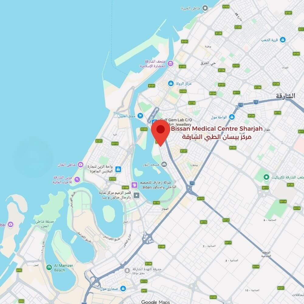
Bissan Medical Centre in Sharjah, Al Majaz 1, provides healthcare services across multiple specialties. To contact us, call +971 (6) 573 7005 | WhatsApp +971 (50) 633 7121 | Email hello@bissanmedical.com.
MOH Approval No. 6MYAJ092-250925